10 Mar 2020
Ticking the boxes – pest control protocols
Emma Gerrard
Job Title
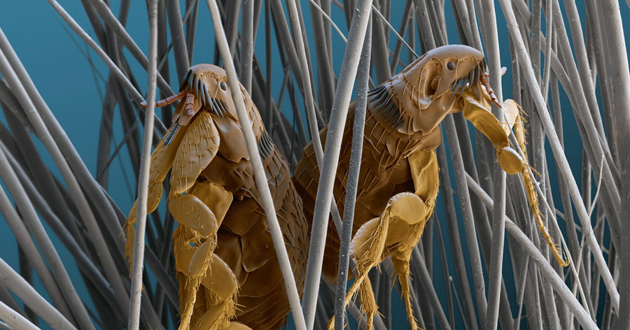
Ctenocphalides felis flea. Image © Bayer Animal Health
The control of parasites in dogs and cats is essential in the prevention of distress, irritation and the spread of vector-borne diseases, some of which are zoonoses.
Routine parasite control forms a vital part of pet health programmes, with some requiring year-round preventive treatment, and others a risk assessment based on geographic and lifestyle factors.
This article aims to give a brief overview of the common pests in cats and dogs.
The control of parasites is essential to maintain the health and welfare of pets, but also to protect public health from zoonotic diseases.
Preventive health care is a huge part of veterinary nursing, and it is important to understand the life cycles and treatment protocols of the main parasite groups to ensure clients and their pets receive the correct advice and products.
VNs are essential in the implementation of individual parasite prevention programmes. A nurse-led parasite consultation can provide individual treatment protocols based on a client’s and pet’s lifestyle, and get key messages across about preventive treatments.
Fleas
Fleas are small wingless insects that form the order Siphonaptera. They are approximately 1mm to 8mm in length, with hindlegs modified for jumping – enabling them to jump up to 200 times their own height. They are external, permanent parasites that are able to complete their life cycle without leaving their host.
Fleas are composed of three sections – the head, thorax and abdomen. More than 2,000 species of fleas exist, but those of importance to domestic pet owners are the cat flea (Ctenocephalides felis) and dog flea (Ctenocephalides canis).
The dog flea is similar in appearance to the cat flea, but much less common. Fleas are haematophagous, and during feeding they inject substances into the skin that results in itching, scratching and skin irritation. Due to their haematophagous nature, fleas can be vectors of various blood-borne diseases.
Fleas prefer warm, humid conditions, and are attracted to a host by their body heat, vibrations and exhaled carbon dioxide. Once on a pet, the eggs that are laid can spread anywhere within the environment, which then act as a source of reinfection when conditions are favourable.
Environmental conditions have a significant impact on the risk of flea infestation. In cool, dry conditions, fleas will remain in their cocoons as pre-emergent adults until they are activated by warmer temperatures and/or host stimuli. Warmer weather causes the pre-emergent adults to hatch, and they will develop quicker in hot and humid weather, with their life cycle being completed in as little as 14 days.
Fleas are highly prevalent on domestic cats and dogs across the UK, with 28.1% of cats and 14.4% of dogs having been demonstrated to be infested (Wright, 2019). Owners should be reminded that a flea infestation can be problematic all year round. Preventive treatment is required during the summer months, but it is equally important during the winter with double-glazed and centrally heated homes.
It is important to remember fleas act as the intermediate host for the tapeworm Dipylidium caninum. Flea larvae ingest D caninum eggs from the environment, and then infested dogs and cats ingest the infected fleas when they groom themselves. It is essential to control fleas to prevent D caninum infection in dogs and cats.
D caninum in fully grown and larval (cystic) stages not only pose a threat to the health of the animal, but also to humans when they act as an accidental host.
Prevention protocols
To instigate effective flea control, the following must be considered:
- multi-animal households
- environmental treatment
- regular vacuuming
- washing bedding
- use of insecticidal sprays
- use of insect development inhibitors and/or insect growth regulators
Ticks
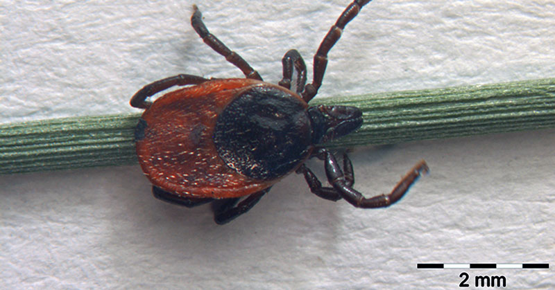
Ticks are arachnids, and, as such, have eight legs, with their bodies composed of two sections (capitulum and Idiosoma).
Ticks are temporary ectoparasites because they spend the majority of their life cycle off the host and only attach for a short period of time to take a blood meal.
Ticks have highly developed mouthparts, which allow them to pierce a hole through the skin, insert their feeding tube and feed from the host. Because of their haematophagous nature, ticks have a remarkable ability to transmit a wide variety of pathogens to animals and humans, compared with any other arthropod vectors.
During the feeding process, the tick injects saliva into the bloodstream, which contains disease-causing organisms, such as bacteria and viruses that have been picked up when feeding on other animals. These organisms enter the bloodstream of the host while the tick is feeding. Ticks have been known as a source of disease for more than 100 years (Swanson et al, 2006).
Unlike permanent parasites, ticks are able to survive on and off the host during all stages of their life cycle. They will only attach on to a host to feed and will feed for extended periods of time. This varies from several days to weeks, depending on factors such as life stage, host type and species. The cuticle (outside surface) of ticks grows to accommodate the large volume of blood ingested, which can be anywhere from 200 to 600 times their unfed bodyweight.
Ticks hatch from their eggs as larvae, with only six legs. These newly hatched larvae look for a host to feed on before moulting to become nymphs. They then feed once more before moulting again to become adults.
While the female tick feeds on the final host, her eggs are fertilised by the male. The female then falls from the host and lays hers eggs in the environment for the life cycle to start over again. Importantly, all life cycle stages can potentially attach to and feed from humans and/or pets.
An exam for ticks should be performed, concentrating on the head, face, legs and ventrum (Wright et al, 2018).
The most common ticks found in the UK are:
- Ixodes ricinus (also known as the castor bean or European sheep tick)
- Ixodes canisuga (host-specific dog tick associated with boarding kennels)
I ricinus is the most common tick species in the UK (Smith et al, 2011) and is commonly referred to as the sheep tick. The risk of dogs being exposed to ticks has increased, with a study having shown nearly one in three dogs were found to host ticks (Abdullah et al, 2016), compared to around one in seven dogs in a previous study (Smith et al, 2011). The risk of exposure to ticks may be seasonal, depending on geographic location.
Less common ticks found in the UK are:
- Ixodes hexagonus (the hedgehog tick)
- Haemaphysalis species
- Dermacentor species
Dermacentor reticulatus is present in endemic foci in the UK. The winter of 2015-16 saw the first recording of a known endemic foci of Babesia canis in the UK (Wright, 2018). Since the outbreaks in Harlow and Romford, data has been gathered regarding the population of Dermacentor ticks, and awareness of the disease has been promoted among veterinary professionals and owners.
Confirmed cases of B canis in dogs that have not travelled abroad have increased the need for surveillance of tick-borne disease in the UK (Swainsbury et al, 2016).
Prevention protocols
Tick prevention must prevent ticks from attaching and taking a blood feed. Tick products will work in a variety of different ways:
- repellents – products containing an ingredient that helps prevent tick attachment
- ingredients that reduce the risk of the tick attaching and taking a blood feed
- acaricide – products that contain an ingredient that kills the tick as it makes contact with the skin
No product is 100% effective at preventing vector-borne pathogens. However, the frequent use of ectoparasiticides may help prevent transmission.
Additional methods of preventing tick-related problems include:
- removing ticks promptly using a tick hook and brush the coat to remove nymphs
- careful grooming and monitoring, especially between March and November
- limiting access to heavily wooded, bushy areas and grassy meadows
Cestodes (tapeworms)
Tapeworms are flat, ribbon-like shapes that are divided into segments, which parasitise the small intestine and absorb nutrients from the final host through the surface of their bodies.
They consist of a scolex (head), which is an attachment organ, a non-segmented neck region and a constantly regenerating series of segments (proglottids). The entire chain of segments (strobila) can grow to several metres long.
The mature tapeworm segments are filled with eggs, and individual segments break off and pass via the dog’s anus into the environment. The neck region constantly generates new segments to replace the mature proglottids that have been shed.
Tapeworms reproduce in intermediate hosts, such as fleas, which act as effective transmitters of tapeworm infestation.
Tapeworms do not cause harm to their hosts unless present in large numbers; however, their treatment is important for hygiene reasons and because some species are zoonotic.
Tapeworms, both in their fully grown and larval (cystic) stages, pose a threat to the health of the animal, but also humans when they act as accidental hosts if eggs are ingested.
The larvae infiltrate the gut wall, and are circulated throughout the body via the blood and the Iymph system. In certain organs of the intermediate host, the larvae develop into an infective cyst. This cyst contains a rudimentary scolex, which may then be ingested by the final host (dog, cat) when they ingest the intermediate host. In the intestinal tract of the final host, the scolex becomes exposed and attaches itself to the intestinal mucosa, where it develops into an adult.
Three main types of cestodes exist:
- Dipylidium canium
- Echinococcus granulosus
- Taenia species
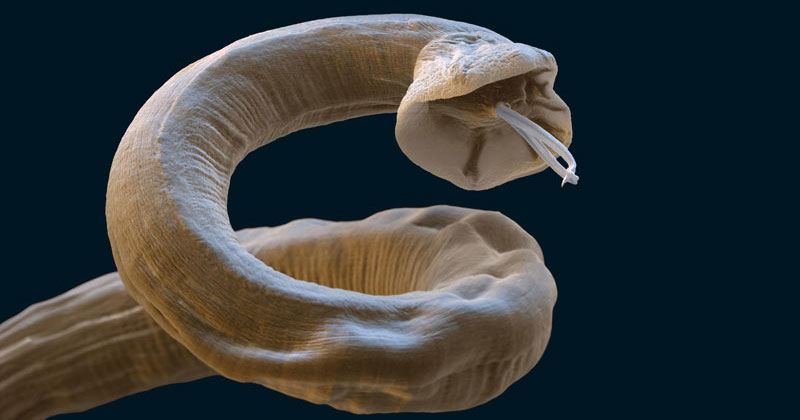
Nematodes (roundworms)
The term roundworm refers to all nematodes, but it can be used to refer specifically to the ascarid group of nematodes:
- Toxocara canis
- Toxocara cati
- Toxascaris leonina
Roundworms are worms of varying sizes that live in the intestine of their host. They attach themselves via their lip-disks to the wall of the intestine, and feed on mucus, blood or intestinal contents.
Most roundworms have a direct life cycle and no intermediate host; although some, such as lungworms, may have one while others can have a paratenic host.
The ingestion of the paratenic host – for example, a mouse – can lead to direct infection of the final host. The larvae frequently travel through the body while they develop, with the final larval stage returning to the intestines, where they metamorphose into adult roundworms and begin egg production.
The common roundworms of dogs and cats are important in companion animal medicine as pathogens, and as a significant zoonotic threat in humans.
Lungworm
Angiostrongylus vasorum (lungworm or French heartworm) is a potentially life-threatening Metastrongylus nematode that inhabits the right ventricle of the heart, and the pulmonary arteries in dogs, foxes and other carnivores. This is in contrast to the rest of the lungworm family, which mainly reside within the bronchi/bronchiolus or the lung itself.
A vasorum should not be confused with Dirofilaria immitis, which also resides in the heart.
The parasite has an indirect life cycle that involves at least two different hosts – gastropod molluscs (intermediate host) and canids (definitive host). The definitive host becomes infected after ingesting the intermediate host – slugs and snails – which carry the lungworm larvae (L3). Infection could also potentially occur after swallowing the slime of an infected slug (Conboy et al, 2017).
It is important to remember molluscs can be extremely small, and dogs can inadvertently ingest them when eating grass, drinking from outdoor puddles and water bowls, or while playing with toys.
Parasite studies have shown A vasorum spreading across the UK, with prevalence in foxes having risen from 7% in 2005 to 18% in 2014 (Morgan et al, 2008; Taylor et al, 2015).
The life cycle of A vasorum is indirect, with dogs/foxes becoming infected when they ingest the intermediate host. The adult worm resides in the right side of the heart and pulmonary arteries of the infected animal.
The female worm then releases eggs, which are carried in the bloodstream to the lung, where they develop into first stage (L1) larvae. The L1 larvae burrow through the walls of the alveoli of the lung, and, from there, are eventually coughed up and swallowed, and passed out into the environment with the faeces, where they can infect slugs and snails.
Further development to L3 takes place within the mollusc. Following ingestion of the mollusc, the larvae are released, subsequently infecting the dog.
The larvae then penetrate the intestinal wall and develop further within the lymph nodes (L5 larvae), which then migrate via the liver and caudal vena cava back to the right side of heart, where development into a new adult worm takes place.
The time taken from ingestion of an infected slug or snail to an adult worm being present in the dog’s heart is, on average, 38 to 57 days.
The diagnosis of canine angiostrongylosis can be challenging due to the wide spectrum of clinical signs. The most common clinical signs of infection are cardiopulmonary, followed by coagulopathies and neurological signs. If left untreated, the dog’s health can rapidly deteriorate and can even result in death.
Treatment and prevention of angiostrongylosis
While it is impossible to stop a dog from eating an infected slug or snail, the products available have been shown to be highly effective at killing the parasite at an early stage and thereby preventing it from completing its life cycle.
A product containing imidacloprid and moxidectin is licensed to prevent angiostrongylosis and patent infection, with 100% efficacy against L4 larvae and immature adults (L5) of A vasorum (Schnyder et al, 2009).
The monthly use of a milbemycin oxime product is indicated for the prevention of angiostrongylosis by reducing levels of infection of L5 and adult parasite stages, with a study having shown a worm count reduction efficacy of 94.9% (Lebon et al, 2016).
Milbemycin oxime is also available in combination with afoxolaner and has been shown to reduce the level of lung pathology associated with angiostrongylosis (Böhm et al, 2014; Lebon et al, 2016). Therefore, vets can provide one treatment rather than recommended a combination of products, or make a choice about which parasite represents the greatest risk in their geographic area – ticks or lungworm.
Protocols for worm prevention
Worming protocols should be tailored to each animal’s individual needs and certain lifestyle factors – such as ectoparasite infection, pet travel and nutrition – may indicate the need for a more regular worming protocol.
The European Scientific Counsel Companion Animal Parasites recommends dogs and cats are wormed every three months with a reputable anthelmintic to ensure they are worm free, and to prevent shedding of eggs into the environment.
Cats that hunt should be wormed on a monthly basis. Breeding/lactating bitches and queens should be wormed accordingly to reduce the risk of infecting puppies and kittens. Puppies and kittens should be wormed from 2 weeks of age, with two-weekly treatments until they are 12 weeks, reducing to monthly treatments until they are 6 months and then three-monthly treatments.
Good personal hygiene should be encouraged, especially for children, and advice should include hand washing after handling, the discouragement of face licking, washing animal bowls separately and ensuring sand pits are covered. Faeces should be removed regularly to reduce environmental contamination with infective parasite stages, and grooming and the examination of pets regularly prevents the risk of coat contamination with worm eggs.
Conclusion
Raising awareness of the health implications of parasites is a massive step towards managing owner compliance and ensuring adequate preventive health care is provided.
VNs in particular are essential to educate clients and increase parasite awareness. Through education of clients on the life cycles of parasites, and the risks they pose to both pets and humans, compliance can be increased.