6 Sept 2022
Surgical management of disc disease in dogs
Bartosz Ropelewski
Job Title
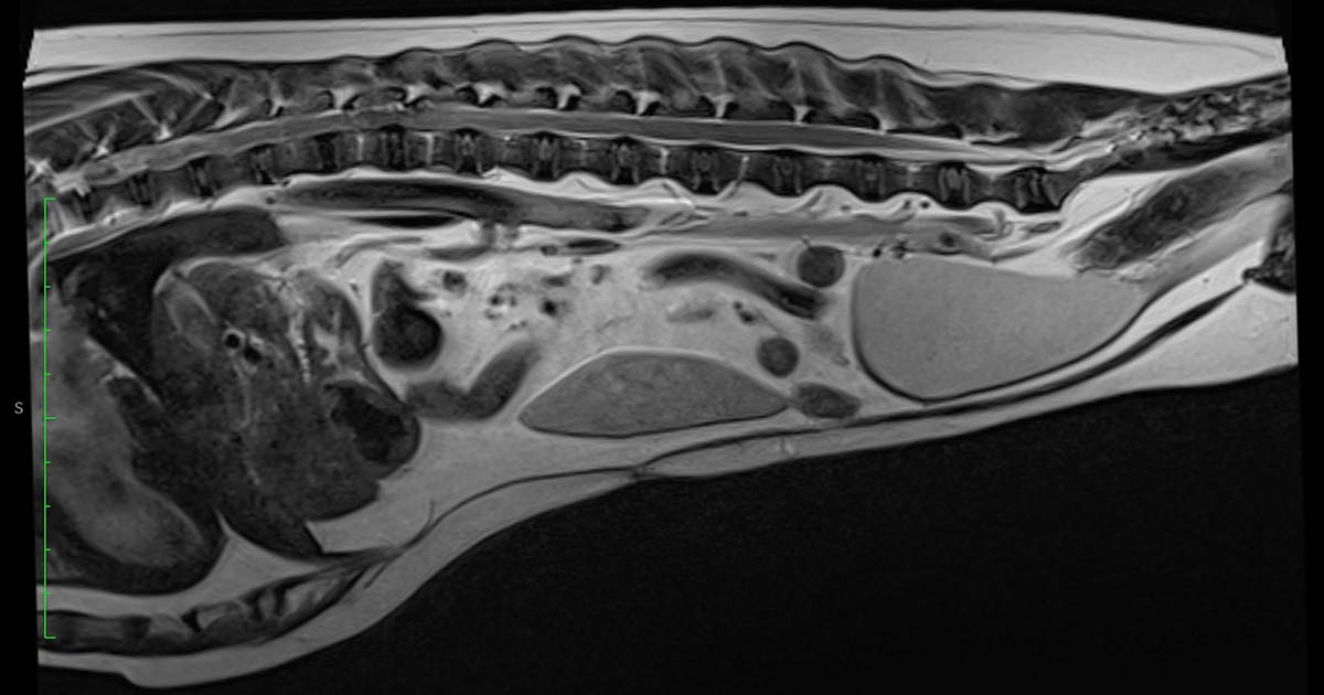
T2-weighted sagittal MRI scan.
A six-year-old dachshund presented for investigation and treatment of a spinal condition. The patient had presented to the first opinion vet a few days earlier with pelvic limb issues. These included evident pelvic limb muscle weakness, proprioceptive positioning deficits, bilateral knuckling and inability to climb stairs.
Initially, the dog was prescribed a course of NSAID medication and gabapentin. The owner reported during this treatment period the dog’s condition became progressively worse.
At presentation to the referral hospital, the patient was not ambulatory and displayed several neurological deficits, including bilateral proprioceptive deficit of the hindlegs, no withdrawal reflexes, no anal reflex, no superficial pain and panniculus reflex deficit from the level of the 13th thoracic vertebrae (T13).
Following discussion of differential diagnoses, possible treatment options, potential complications and prognosis with the owner, the patient was admitted for further investigation.
MRI scan was performed under general anaesthetic. Premedication was with medetomidine 0.005mg/kg IV and methadone 0.3mg/kg IV. The patient was induced with 4mg/kg propofol IV to effect and intubated with a 6mm cuffed endotracheal tube. Anaesthesia was maintained on sevoflurane and oxygen.
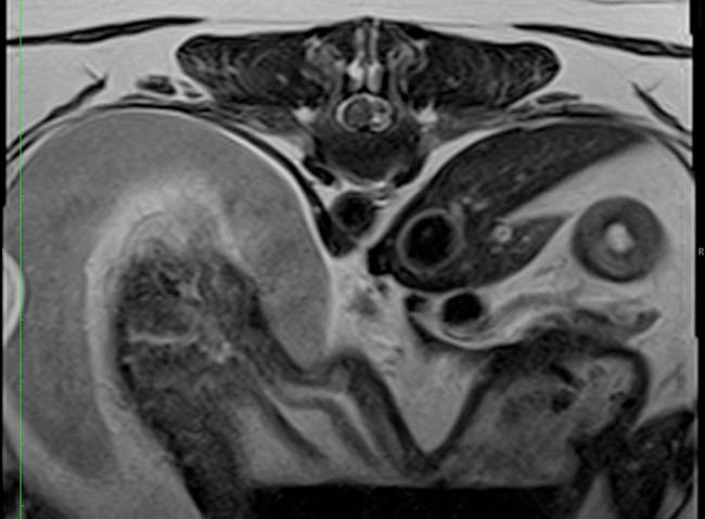
The MRI scan showed a right sided extrusion of the T12-T13 intervertebral disc, causing an evident spinal cord compression.
After consultation with the owner, the decision was made to perform surgical spinal cord decompression. Cefuroxime 15mg/kg IV and paracetamol 10mg/kg IV were given. A constant rate infusion of ketamine was given throughout general anaesthetic at a rate of 0.6ml/hr, reduced to 0.3ml/hr on recovery. IV fluids at the surgical rate 5ml/kg were given throughout surgery.
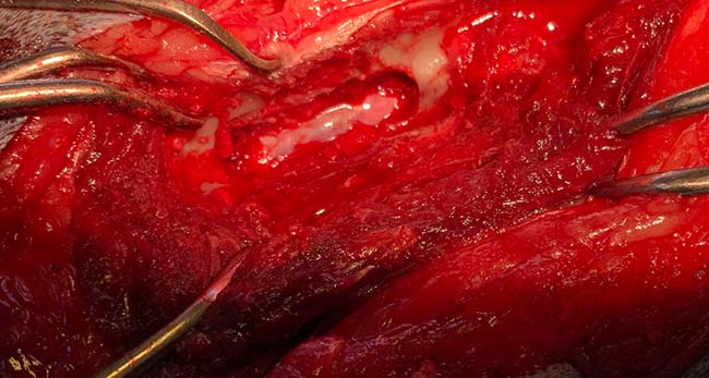
A dorsal midline incision was performed. The multifidus musculature was elevated with the use of the periosteal elevator. This exposure allowed sufficient elevation of the tendinous epaxial muscle attachments to help visualise the lateral aspect of the spinous process, lamina and facet joint up to the level of accessory process.
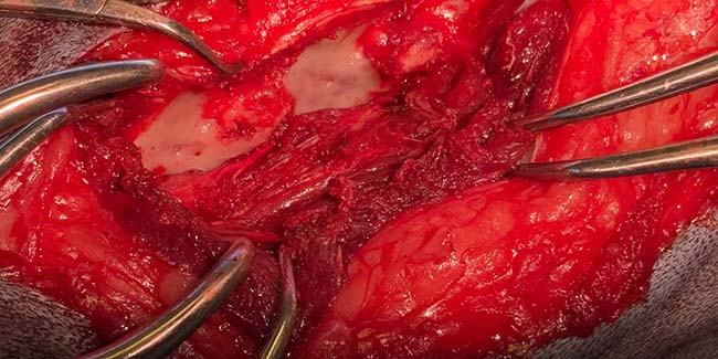
A right-sided T12-T13 hemilaminectomy was performed. The hemilaminectomy was completed using a surgical burr and lempert rongeurs. The spinal nerve was identified and retracted with a spinal hook.
A urinary catheter was placed into the bladder after completing the surgical procedure. The patient recovered from anaesthesia uneventfully. The patient had intensive physiotherapy during the period of hospitalisation. The urinary catheter was removed and spontaneous urination restored. The dog gradually improved and was discharged to the owner.
At discharge, the patient was ambulatory and had mild proprioceptive deficits on the right hindleg. The patient underwent physiotherapy and hydrotherapy.
The dog was reassessed after four weeks; at this time, he had a normal gait and posture with no obvious neurological deficits.
Discussion
Chondrodystrophic breed (CD) dogs are predisposed to intervertebral disc disease (IVDD) due to their age and breed-related metaplastic degeneration of the discs (Bergknut et al, 2011).
The process of metaplastic disc degeneration occurs much faster in CD dogs compared to non-CD dogs (NCD; Hansen et al, 2017).
Multiple studies confirm that the process of disc degeneration starts in the central nucleus pulposus (Braund et al, 1975; Ghosh et al, 1975).
Hansen, in his 1952 study, distinguished two different pathophysiological processes effecting IVDD in CD dogs, calling this process “chondroid metaplasia” and, respectively in NCD breeds, “fibroid metaplasia” (Hansen, 1952).
A more recent study comparing intervertebral disc degeneration affecting CD and NCD dogs confirmed the same type of histopathological changes, called chondroid metaplasia. Histopathological disc assessment did not confirm presence of fibroblasts and fibrocyte-like cells within the degenerated nucleus pulposus of the affected discs in both NCD and CD dogs (Hansen et al, 2017). During the process of disc degeneration (chondroid metaplasia), a significant decrease of proteoglycans (chondroitin sulfate), glycosaminoglycans and water occurs, as well as increased levels of collagen (Ghosh et al, 1977; Bergknut et al, 2012). This affects the ability of the disc to absorb shock by causing damage to the annulus fibrosus and progressive endplate sclerosis (Hansen, 1952).
Types of intervertebral disc herniation
Disc decompression techniques
Depending on the location and type of disc, different types of spinal cord decompression may be selected.