18 Mar 2019
Case of dog with severe anaemia
Francesco Cian presents the case of an adult male cocker spaniel with a clinical history including lethargy and anorexia in his latest Haematology Hub.
Francesco Cian
Job Title
Figure 1.Wright-Giemsa 50×.
The image (Figure 1, Wright-Giemsa 50×) and data (Table 1) are from an ethylenediamine tetra-acetic acid (EDTA) blood sample of an adult male cocker spaniel with a clinical history of a week of lethargy, anorexia, sporadic episodes of vomiting and pale mucous membranes.
Based on the data and blood smear picture, try to answer the following questions.
What is the most likely explanation for the severe anaemia?
There is evidence of a severe anaemia that is macrocytic (high mean corpuscular volume) and hypochromic (low mean corpuscular haemoglobin concentration). This is due to the presence of early red blood cells (polychromatophils or reticulocytes) released by the bone marrow in response to an increased loss, or destruction of, red blood cells in the peripheral circulation.
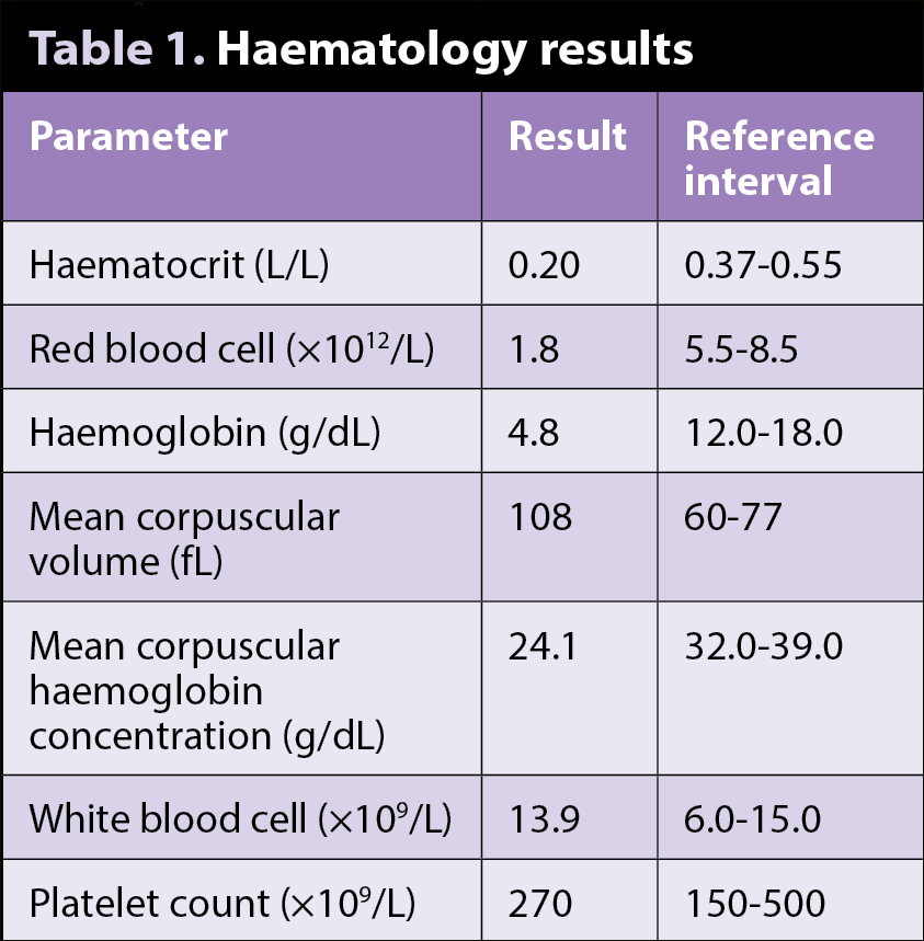
Polychromatophils can be appreciated on the blood smear (blue arrows), as red blood cells are larger than normal and blue-purple in colour. This is due to the presence of residual RNA, which offsets the red of haemoglobin, imparting a blue-purple colour to the cells. A few nucleated red blood cells, called rubricytes (purple arrow), are also noted.
Given this evidence of active haematopoiesis, the anaemia is considered regenerative, and may be due to blood loss and/or haemolysis. In this specific case, no clinical evidence of blood loss occurred. Moreover, on blood smear examination, variable numbers of red blood cells are observed – smaller than normal, with dense staining properties and no central pallor.
These cells are called spherocytes (red arrows), and are often seen in immune-mediated haemolytic anaemia (IMHA) cases. Formation of spherocytes results when antibody-coated red blood cells bind to macrophages – resulting in partial phagocytosis of the red blood cell. The remnant has a reduced surface area to volume ratio and assumes a sphere shape rather than a disc.
Which other diagnostic tests would you suggest to confirm these findings?
A saline agglutination test and Coombs test. The latter is a supportive diagnostic test for IMHA and aims to identify the presence of antibodies (IgG, IgM) or complement (C3) on red blood cells. Both  false-positive and false-negative results may occur; therefore a diagnosis of IMHA should not rely on the results of the Coombs test alone.
Which are the most common causes of immune-mediated haemolytic anaemia in dogs?
IMHA in dogs is most commonly a primary idiopathic condition, with autoantibodies produced against the animal’s own red blood cell membrane antigens. In these forms, a genetic susceptibility component is likely to exist, as evidenced by the high prevalence in certain breeds, such as cocker spaniels.
IMHA may also be secondary and triggered by an inflammatory or neoplastic process, which induces an immunologic response to non-self antigens that have modified, or are associated with, normal red blood cell membranes.
Tick-borne infectious diseases have also been linked to immune-mediated disease – for example, Babesia species, Ehrlichia species or haemotropic Mycoplasma species in splenectomised or immunosuppressed dogs.
Latest news
