12 Jan 2021
Allergies in dogs: how to reach a diagnosis part 2
Diana Ferreira
Job Title
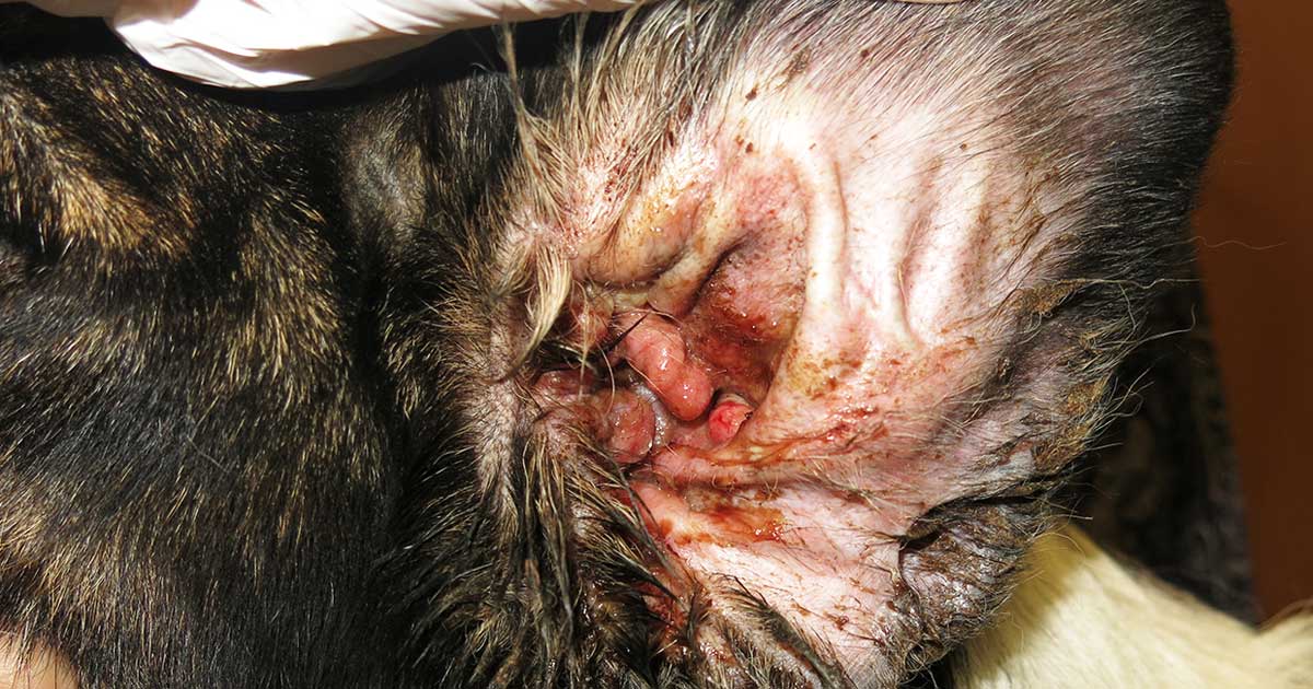
Figure 1. Otitis externa in an allergic dog.
Having excluded parasitic causes and addressed secondary infections, if the animal maintains the pruritus, and the clinical signs and lesion distribution are compatible, an allergic dermatitis is diagnosed. The next step is to investigate and explore allergic causes.
The most important clinical features of allergies include:
- The presence of pruritus.
- Associated primary skin lesions consisting of erythematous macules or patches and small papules.
- A characteristic distribution affecting the face, antecubital fossae, interdigital skin, ventral abdomen, perineum, the ventral aspect of the proximal tail and the ear canals, with otitis externa occurring frequently (Figure 1).
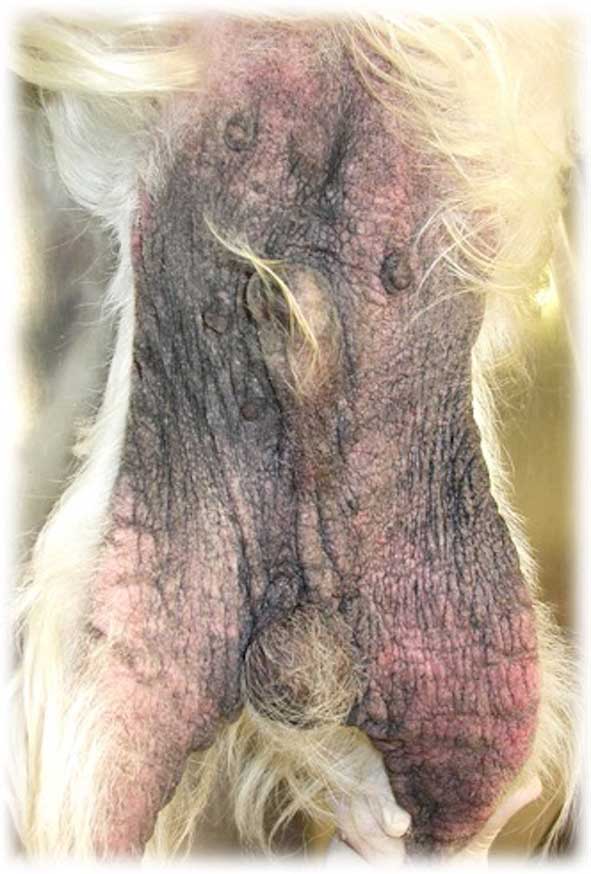
- In chronically affected animals, in addition to the primary lesions, excoriations, self-induced alopecia, lichenification and hyperpigmentation may occur (Figure 2).
Because atopic dermatitis is a diagnosis of exclusion, the first step is to look at food hypersensitivity.
Food allergy
The influence of food allergens should be determined in all cases of non-seasonal pruritus.
Currently, performing an elimination trial and a subsequent provocation test represents the most reliable method by which to reach a diagnosis of food allergy. Serological and skin tests are considered inaccurate in the diagnosis of food allergy; therefore, they are currently not recommended.
An elimination diet consists of feeding exclusively a source of protein and carbohydrate that the animal never ate before. The dietary history of the patient will have to be taken into account when choosing a suitable diet.
For the elimination trial, three types of diets could be used.
Home-cooked diet
A home-cooked diet is considered the most reliable one for the diagnosis. A better control exists of the ingredients included in the diet, and any problems that could occur related to the manufacturing of a commercial diet are eliminated.
Novel foods that can be used include filleted white fish, canned tuna in water, rabbit, venison, turkey, rice, potatoes, ostrich, yams, and pinto beans.
Hypoallergenic commercial diets
Hypoallergenic commercial diets are based on hydrolysed proteins. These are very practical for owners; however, they may maintain some persistent allergenicity.
Consideration should be given to the source of ingredients when using hydrolysed protein diets in elimination trials because antigenic sites may not be fully destroyed.
Novel protein commercial diets
Various brands offer novel proteins such as duck, lamb, venison, rabbit, white fish or salmon. One study has shown contamination of commercial foods with other food allergens not listed on the ingredient label, which can represent a concern.
The elimination diet should be carried out for a minimum of eight weeks and clients should be educated about the importance of strict adherence to the food trial. No other food, flavoured toys or treats should be allowed during the trial. Chewable medications should also be discontinued.
The dog’s clinical signs – including pruritus, inflammation and/or the development of recurrent infections – should be monitored during the food trial. If at the end of the food trial the clinical signs resolved or improved, the next step is to challenge the dog with the previous diet to determine if the animal again relapses.
It is important that the challenge is performed to all dogs that improve. In many cases an improvement can occur as a result of treatments, resolution of infections, flea control, a superior quality diet or changes of season for dogs that have atopic disease, and not because the animal is food allergic. If the animal is food allergic, it will typically relapse with clinical signs within 1 or 2 days, and as long as 7 to 14 days after re-challenge.
For diagnosis, a clinical improvement has to occur during the food trial, with a worsening of clinical signs on a challenge with the previous diet, and again an improvement when the hypoallergenic diet is subsequently reintroduced.
The most common food allergens in dogs are beef, dairy products, chicken and wheat.
If a reduction in the pruritus is not obtained with the food trial – and a typical clinical presentation, lesion distribution and history exists – the diagnosis of atopic dermatitis is made.
Atopic dermatitis
The diagnosis of canine atopic dermatitis (AD) is based on clinical signs and made by exclusion. No need exists to perform complementary tests – such as intradermal or serological tests – to obtain a diagnosis; however, these tests may help in the clinical and therapeutic decision-making.
Favrot et al (2010) proposed a number of clinical criteria for the diagnosis of canine AD (Panel 1). In the presence of five of the eight presented criteria, a sensitivity and specificity around 80% is obtained. These criteria, however, are used essentially for clinical trials and do not replace detailed diagnostic work. Their use as the only method of diagnosis would misdiagnose 20% of patients.
1. Age at onset less than three years.
2. Mostly indoor.
3. Corticosteroid-responsive pruritus.
4. Chronic or recurrent yeast infections.
5. Affected front feet.
6. Affected ear pinnae.
7. Non-affected ear margins.
8. Non-affected dorso-lumbar area.
Serological or intradermal tests should only be used to confirm the clinical diagnosis of canine AD and should not be considered as a screening test for obtaining an AD diagnosis.
Intradermal tests and allergen-specific IgE serology still lack standardisation, and it is known that false positive and false negative results do occur. Between 10% and 30% of dogs with a clinically confirmed canine AD may show a negative intradermal skin test. This can occur due to several factors. Among them are:
- improper technique
- low concentration of the allergens used for the test
- drug interference
- factors related to the animal itself, such as stress
- incorrect selection of the allergens for the test
- test performed too long after the peak of the allergy season, or too long after clinical signs have disappeared
- the animal being affected by atopic-like dermatitis (clinically identical to canine AD; however, IgE response to environmental or other allergens cannot be documented)
Serological and intradermal skin tests can, however, be used to:
- document whether the disease is associated with allergen-specific IgE
- implement avoidance techniques to the allergens detected in the tests
- select the allergens to be included in an allergen-specific immunotherapy for desensitisation treatment
When are skin biopsies considered in the diagnostic work?
Biopsies can be useful when we need to exclude diseases such as cutaneous epithelial T-cell lymphoma. In cases of allergic dermatitis, biopsy findings are usually non-specific and inconclusive regarding the cause of the allergic disease.
In conclusion
The diagnosis of allergic dermatitis in dogs is always often challenging. In part, because of the absence of pathognomonic clinical signs in these diseases and because of the potential variability in the clinical signs among affected animals.
A comprehensive, systematic diagnostic workup is recommended to give the best chance of obtaining the correct diagnosis.